The material stays in the sentence
GHK-Cu wound research is also materials research
The peptide often arrives inside a gel, a scaffold or a modified fibre. Removing that material from the headline removes part of the intervention.
Stations
Over 95%, inside a particular mouse experiment
The 2025 infected-wound study tested a food-derived tripeptide-copper composite hydrogel. In mice with infected full-thickness wounds, it achieved over 95% closure by day 12, compared with about 65% in controls. The work also found antimicrobial activity against S. aureus and E. coli, lower IL-6 and TNF-alpha inflammatory signals, more collagen deposition and new blood vessels.
It is an impressive bundle of results for that experimental material. I’m reluctant to give the peptide sole credit for a composite intervention, especially when wound closure, bacterial control and tissue organization are being pursued together. A gel is doing more than carrying a convenient ingredient name.
The result came from mouse wounds and associated laboratory work. It does not establish a human wound treatment, a scar-fading result or a reason to put GHK-Cu on an injury.
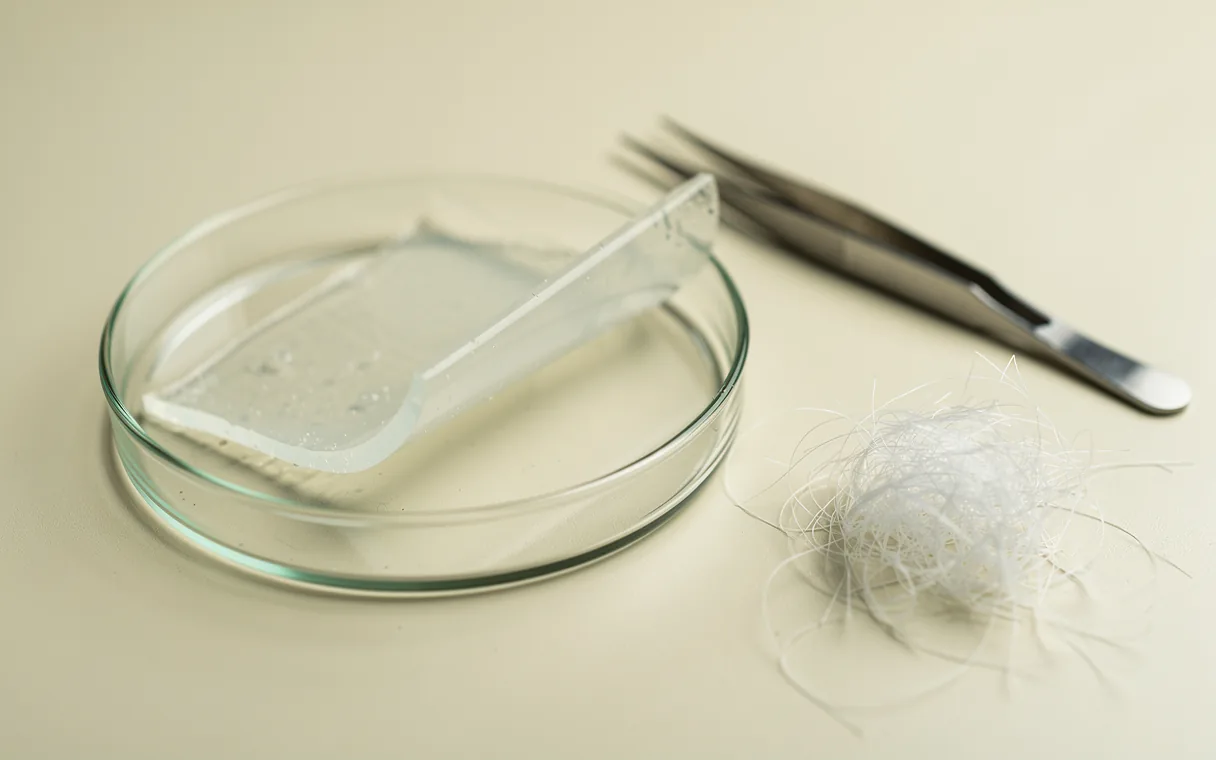
The carrier has a job description
In the hyaluronic-acid hydrogel study, GHK peptide nanofibers were embedded in a material that could be crosslinked with light. The experiments included human dermal fibroblasts in culture and mouse wound models; they found accelerated closure, denser remodeled collagen and increased blood-vessel formation. This was a modified peptide-and-material system.
The earlier GHK-modified alginate work asked a different question. Human stem/stromal cells grown with the modified gel released more growth factors, and the cell signals supported vessel-lining cell proliferation and tube formation in culture. The culture test measured tube formation; circulation in patients was outside its scope.
The materials are the bit I’d investigate further: they make the intervention tangible. Where the peptide sits, how it is attached and what surrounds it are part of the experiment. Treating all of this as a single finding about loose GHK-Cu would erase the engineering.
GHK-Cu scaffold coating: cell viability and bacteria
The coated-scaffold study used a 1 mM GHK-Cu coating solution on a poly(epsilon-caprolactone), collagen and chitosan scaffold. Human dermal fibroblast viability was higher after three days than on uncoated controls, and antibacterial activity against E. coli and S. aureus appeared within one hour. Those times describe the cell and microbiological experiments; treatment schedules remain unestablished.
The 2008 tissue-remodeling review gathers a broader pattern of collagen production, repair-cell recruitment and blood-vessel growth. That pattern helps explain the wound interest. It cannot replace a direct human comparison for a particular material or outcome.
Better-organized collagen in the animal and laboratory work is relevant to scar research, but it has not established that GHK-Cu fades human scars. The next experiment I’d welcome is specific: the material itself, the relevant wound type and a human endpoint, all in the same sentence.